
If your dog has had a suspected bad reaction to a medication, imaging is usually about looking for something else that could explain the signs: fluid in the lungs, a blockage in the gut, a mass, or a change in the brain. An X-ray gives a flat shadow picture of dense tissue, a CT scanner builds a slice from many angles around the body, and an MRI uses magnetic fields and radio waves with no ionising radiation at all. The choice belongs to your veterinarian, who weighs the dog’s condition, the suspected drug, and what question the image needs to answer.
What is an X-ray image and why is it like a shadow?
An X-ray is a two-dimensional picture made by passing a beam of X-ray photons through the body onto a detector on the far side. Dense material such as bone stops more of the beam, so less reaches the detector and that area looks white. Air-filled lung tissue stops very little, so more reaches the detector and that area looks black. Soft tissues fall in between, in overlapping shades of grey.
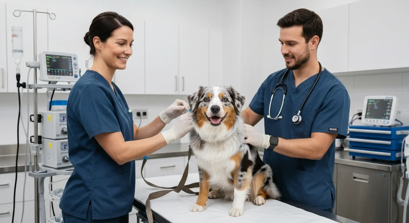
That is why people describe it as a shadow. The image is a projection, not a slice. Everything along the path of the beam is flattened onto one plane, so a rib sits on top of a lung field, and a stomach sits on top of a spine. A radiographer usually takes at least two views, often one from the side and one from above, because a single projection can hide a lesion behind another structure. For a dog with a suspected drug reaction, a chest X-ray is a common first step when the vet wants to know whether there is fluid, an infection, or a change in heart size.
A useful way to picture the difference between projection and slice is to think of a loaf of bread. An X-ray is the shadow the whole loaf casts on a wall. A CT slice is one cut slice laid flat on a board. If you want to understand how that shadow is produced and read, a plain-language walkthrough of the X-ray image, shadow principle is a sensible place to start, because the same physics sits behind every projection image taken in a veterinary practice or a hospital.
How does a CT scanner create a computed slice?
CT stands for computed tomography. The scanner does not take one picture from one direction. It rotates an X-ray tube and a detector array around the patient and takes hundreds of readings from many angles. A computer then reconstructs those readings into cross-sectional slices, each one a thin disc through the body.
The word computed is doing real work here. The machine is not photographing a slice; it is calculating one from the pattern of how much radiation reached the detectors at each angle. Modern scanners can produce slices a millimetre or less thick, and the software can stack them into views from the side, from the front, or in three dimensions. That is why CT shows a small nodule, a fracture, or a bleed that a plain X-ray might blur into the surrounding tissue.
For a dog, CT usually means general anaesthesia or heavy sedation, because the animal has to lie still while the gantry spins. The scan itself is short, often a matter of seconds to a couple of minutes, but the preparation and recovery take longer. Contrast agents may be injected to make blood vessels or certain organs stand out, and the veterinary team will check kidney values beforehand because contrast is cleared by the kidneys.
Why does an MRI scan not use ionising radiation?
MRI stands for magnetic resonance imaging. It uses a strong magnetic field and pulses of radio waves, not X-rays. The hydrogen nuclei in water and fat inside the body behave like tiny compass needles; the magnetic field lines them up, the radio pulses knock them out of alignment, and the signal they give off as they relax is turned into an image. No ionising radiation is involved at any point.
That matters for repeated imaging, for young animals, and for anyone who is pregnant or has had a lot of scans. It does not make MRI risk-free. The strong magnetic field means metal implants, some collars, and certain medical devices are a safety concern, and the scanner is loud enough that ear protection is needed. MRI also takes longer than CT, often twenty to sixty minutes, and motion ruins the pictures, so anaesthesia is almost always required in dogs.
MRI is particularly good at soft tissue: the brain, the spinal cord, joints, and some abdominal organs. If a dog has neurological signs after a drug reaction, such as tremors, seizures, or a changed gait, the vet may want MRI to look at the brain or spine rather than CT or X-ray.
What happens between the request and the report?
Imaging is a pathway, not a single event. A clinician writes a request explaining the question to be answered, for example whether there is free fluid in the abdomen or a lesion in the brain. The imaging service checks that request against safety rules before the appointment, including whether the examination is justified and whether the dose or the magnetic field exposure has been optimised for the patient.
On the day, the radiographer or radiologist positions the patient, sets the machine, and may give contrast. After the scan, the images are reviewed and a written report goes back to the referring vet. In the UK, the relevant framework for X-ray and CT safety is the Ionising Radiation (Medical Exposure) Regulations 2017, usually shortened to IR(ME)R 2017, which sets out who can justify, authorise, and perform exposures. MRI sits outside those regulations because it does not use ionising radiation, but it has its own safety checks.
Does my dog need imaging at all after a reaction?
Not always. Many suspected drug reactions are managed by stopping the drug, supporting the dog, and monitoring. Imaging is added when the clinical picture suggests something structural: breathing that does not improve, a distended abdomen, pain, neurological signs, or a fever that will not settle. The vet decides, and the decision is based on the individual dog, not on a protocol.
If imaging is advised, ask what question it is meant to answer and what will change depending on the result. That conversation often matters more than the choice of machine. A normal scan can be as useful as an abnormal one, because it rules out a problem and lets the team focus on the reaction itself.
How do I prepare my dog and myself?
Follow the fasting instructions exactly if anaesthesia is planned. Tell the team every medication, supplement, and herbal product your dog takes, including the one you suspect caused the reaction, and say when the last dose was given. Mention any metal your dog carries, such as a microchip is fine but a metal tag or a surgical implant is not, and flag any previous reaction to contrast.
Bring the MDR1 test result if your dog has one. It does not change the physics of the scanner, but it changes which sedatives and anaesthetic drugs are safe, and that is a conversation for the veterinarian. Ask who will read the images and when the report is expected, so you know when to call for results.
After the appointment, keep the discharge notes with the rest of your dog’s records. If a reaction is confirmed or suspected, the details of what was given, when, and how the dog responded are the most useful thing you can hand to the next vet who sees your dog.